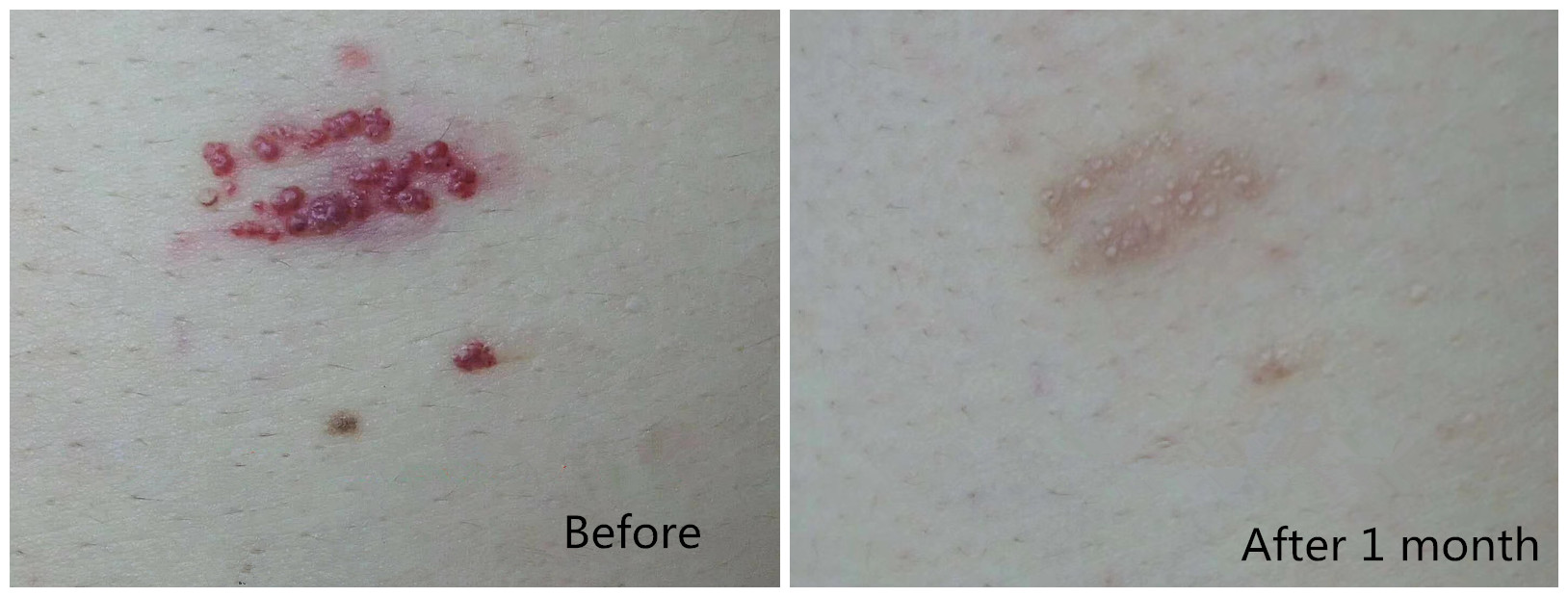
Long-pulsed 1064 Nd:YAG laser proves to be an effective treatment for hemangioma and vascular malformation in darker skin patients with its major advantages of being a safe, well-tolerated, cost-effective procedure with minimal downtime and minimal side effects.
Laser treatment of superficial and deep leg veins as well as various other vascular lesions remains one of the more common applications of lasers in dermatology and phlebology. In fact, lasers have largely become the treatment of choice for vascular birthmarks such as hemangiomas and port-wine stains and the definitive treatment of rosacea. The range of congenital and acquired benign vascular lesions effectively treated with lasers continues to expand and is described by the principle of selective photothermolysis. In the case of vascular specific laser systems, the intended target is intravascular oxyhemoglobin.
By targeting oxyhemoglobin, energy is transferred to the surrounding vessel wall. Currently, the 1064-nm Nd: YAG laser and the visible/near infrared (IR) intense pulsed light (IPL) devices both give good results. The main difference, however, is that Nd: YAG lasers can penetrate much deeper and are therefore more suitable for the treatment of larger, deeper blood vessels such as leg veins. Another advantage of the Nd: YAG laser is its lower absorption coefficient for melanin. With a lower absorption coefficient for melanin, there is less concern for collateral epidermal damage so it may be more safely used to treat darker pigmented patients. The risk for post inflammatory hyper pigmentation can further be minimized by epidermal cooling devices. Epidermal cooling is imperative to safeguard against collateral damage from melanin absorption.
Leg vein therapy is one of the most commonly requested cosmetic procedures. Ecstatic venules present in approximately 40% of women and 15% of men. More than 70% have a family history. Often, pregnancy or other hormonal influences are implicated. Although a primarily cosmetic problem, more than half of these vessels may become symptomatic. The vascular network is a complex system of multiple vessels of different caliber and depths. Venous drainage of the leg consists of two primary channels, the deep muscular plexus and the superficial cutaneous plexus. The two channels are connected by deep perforating vessels. Smaller cutaneous vessels, which reside in the upper papillary dermis, drain to deeper reticular veins. The larger reticular veins dwell in the reticular dermis and subcutaneous fat. The superficial veins may be as large as 1 to2 mm. Reticular veins may be 4 to 6 mm in size. Larger veins have thicker walls, have a higher concentration of deoxygenated blood, and can be more than 4 mm deep. Variations in vessel size, depth, and oxygenation influence modality and efficacy of leg vein therapy. Visible light devices targeting the oxyhemoglobin absorption peaks may be acceptable for treating very superficial telangiectasias on the legs. Longer-wavelength, near-IR lasers allow deeper penetration of the tissue and may even be used to target deeper reticular veins. Longer wavelengths also heat more uniformly than the shorter wavelengths with higher absorption coefficients.
Laser leg vein treatment end points are immediate vessel disappearance or visible intravascular thrombosis or rupture. Microthrombi may be appreciable in the vessel lumen. Likewise, perivascular extravasations of blood may be apparent from vessel rupture. Occasionally, an audible pop may be appreciated with rupture. When very short pulse durations, less than 20 milliseconds, are used, spot sized purpura may occur. This is likely secondary to rapid microvascular heating and rupture.
The Nd: YAG modifications with variable spot sizes (1-6 mm) and higher fluences allow for focal vascular elimination with more limited collateral tissue damage. Clinical evaluation has shown that pulse durations between 40 and 60 milliseconds provide optimal treatment of leg veins.
The most common adverse side effect of laser treatment of leg veins is post inflammatory hyper pigmentation. This is seen more commonly with darker skin types, sun exposure, shorter pulse durations (<20 milliseconds), ruptured vessels, and vessels with thrombus formation. It fades with time, but this may be a year or longer in some cases. If excessive heating is delivered by either inappropriate fluence or pulse duration, ulceration and subsequent scarring may ensue.
Post time: Oct-31-2022